


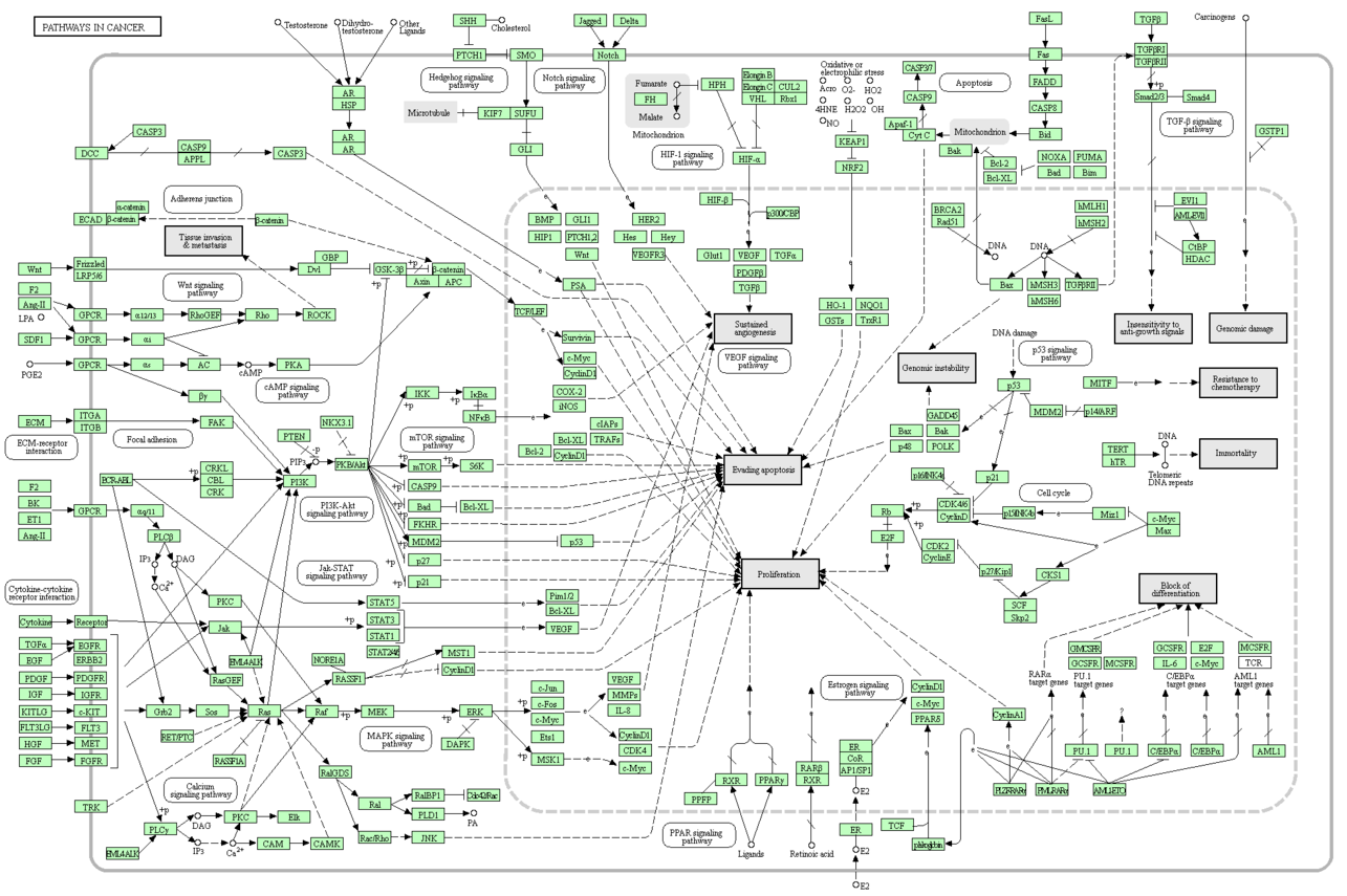
1.肿瘤疾病相关通路
2.各个通路详细介绍
- 1.MAPK (ERK) signaling
-
https://www.genome.jp/dbget-bin/www_bget?pathway:map04010
The mitogen-activated protein kinase (MAPK) cascade is a highly conserved module that is involved in various cellular functions, including cell proliferation, differentiation and migration. Mammals express at least four distinctly regulated groups of MAPKs, extracellular signal-related kinases (ERK)-1/2, Jun amino-terminal kinases (JNK1/2/3), p38 proteins (p38alpha/beta/gamma/delta) and ERK5, that are activated by specific MAPKKs: MEK1/2 for ERK1/2, MKK3/6 for the p38, MKK4/7 (JNKK1/2) for the JNKs, and MEK5 for ERK5. Each MAPKK, however, can be activated by more than one MAPKKK, increasing the complexity and diversity of MAPK signalling. Presumably each MAPKKK confers responsiveness to distinct stimuli. For example, activation of ERK1/2 by growth factors depends on the MAPKKK c-Raf, but other MAPKKKs may activate ERK1/2 in response to pro-inflammatory stimuli.
- 2.RAS signaling
-
https://www.genome.jp/dbget-bin/www_bget?pathway:map04014
The Ras proteins are GTPases that function as molecular switches for signaling pathways regulating cell proliferation, survival, growth, migration, differentiation or cytoskeletal dynamism. Ras proteins transduce signals from extracellular growth factors by cycling between inactive GDP-bound and active GTP-bound states. The exchange of GTP for GDP on RAS is regulated by guanine nucleotide exchange factors (GEFs) and GTPase-activating proteins (GAPs). Activated RAS (RAS-GTP) regulates multiple cellular functions through effectors including Raf, phosphatidylinositol 3-kinase (PI3K) and Ral guanine nucleotide-dissociation stimulator (RALGDS).
- 3.PI3K signaling
-
https://www.genome.jp/dbget-bin/www_bget?pathway:map04151
The phosphatidylinositol 3' -kinase(PI3K)-Akt signaling pathway is activated by many types of cellular stimuli or toxic insults and regulates fundamental cellular functions such as transcription, translation, proliferation, growth, and survival. The binding of growth factors to their receptor tyrosine kinase (RTK) or G protein-coupled receptors (GPCR) stimulates class Ia and Ib PI3K isoforms, respectively. PI3K catalyzes the production of phosphatidylinositol-3,4,5-triphosphate (PIP3) at the cell membrane. PIP3 in turn serves as a second messenger that helps to activate Akt. Once active, Akt can control key cellular processes by phosphorylating substrates involved in apoptosis, protein synthesis, metabolism, and cell cycle.
- 4.WNT signaling
-
https://www.genome.jp/dbget-bin/www_bget?pathway:map04310
Wnt proteins are secreted morphogens that are required for basic developmental processes, such as cell-fate specification, progenitor-cell proliferation and the control of asymmetric cell division, in many different species and organs. There are at least three different Wnt pathways: the canonical pathway, the planar cell polarity (PCP) pathway and the Wnt/Ca2+ pathway. In the canonical Wnt pathway, the major effect of Wnt ligand binding to its receptor is the stabilization of cytoplasmic beta-catenin through inhibition of the bea-catenin degradation complex. Beta-catenin is then free to enter the nucleus and activate Wnt-regulated genes through its interaction with TCF (T-cell factor) family transcription factors and concomitant recruitment of coactivators. Planar cell polarity (PCP) signaling leads to the activation of the small GTPases RHOA (RAS homologue gene-family member A) and RAC1, which activate the stress kinase JNK (Jun N-terminal kinase) and ROCK (RHO-associated coiled-coil-containing protein kinase 1) and leads to remodelling of the cytoskeleton and changes in cell adhesion and motility. WNT-Ca2+ signalling is mediated through G proteins and phospholipases and leads to transient increases in cytoplasmic free calcium that subsequently activate the kinase PKC (protein kinase C) and CAMKII (calcium calmodulin mediated kinase II) and the phosphatase calcineurin.
- 5.NOTCH signaling
-
https://www.genome.jp/dbget-bin/www_bget?pathway:map04330
The Notch signaling pathway is an evolutionarily conserved, intercellular signaling mechanism essential for proper embryonic development in all metazoan organisms in the Animal kingdom. The Notch proteins (Notch1-Notch4 in vertebrates) are single-pass receptors that are activated by the Delta (or Delta-like) and Jagged/Serrate families of membrane-bound ligands. They are transported to the plasma membrane as cleaved, but otherwise intact polypeptides. Interaction with ligand leads to two additional proteolytic cleavages that liberate the Notch intracellular domain (NICD) from the plasma membrane. The NICD translocates to the nucleus, where it forms a complex with the DNA binding protein CSL, displacing a histone deacetylase (HDAc)-co-repressor (CoR) complex from CSL. Components of an activation complex, such as MAML1 and histone acetyltransferases (HATs), are recruited to the NICD-CSL complex, leading to the transcriptional activation of Notch target genes.
- 6.HH signaling
-
https://www.genome.jp/dbget-bin/www_bget?pathway:map04340
The Hedgehog (Hh) signaling pathway has numerous roles in the control of cell proliferation, tissue patterning, stem cell maintenance and development. The primary cilium is an important center for transduction of the Hedgehog signal in vertebrates. In Hh's absence, the Ptch receptor localizes to the cilium, where it inhibits Smo activation. Gli proteins are phosphorylated by PKA, CKI and GSK3B and partially degraded into truncated Gli repressor form (GliR) that suppresses Hh target gene transcription in the nucleus. In Hh's presence, Ptch disappears from the cilium, and activated Smo contributes to the translocation of the protein complex Gli, Sufu, Kif7 to ciliary tip, where Gli dissociates from the negative regulator Sufu. The production of Gli activator form (GliA) occurs and the increased nuclear accumulation of GliA results in activation transcription of Hh target genes.
- 7.TGFB signaling
-
https://www.genome.jp/dbget-bin/www_bget?pathway:map04350
The transforming growth factor-beta (TGF-beta) family members, which include TGF-betas, activins and bone morphogenetic proteins (BMPs), are structurally related secreted cytokines found in species ranging from worms and insects to mammals. A wide spectrum of cellular functions such as proliferation, apoptosis, differentiation and migration are regulated by TGF-beta family members. TGF-beta family member binds to the Type II receptor and recruits Type I, whereby Type II receptor phosphorylates and activates Type I. The Type I receptor, in turn, phosphorylates receptor-activated Smads ( R-Smads: Smad1, Smad2, Smad3, Smad5, and Smad8). Once phosphorylated, R-Smads associate with the co-mediator Smad, Smad4, and the heteromeric complex then translocates into the nucleus. In the nucleus, Smad complexes activate specific genes through cooperative interactions with other DNA-binding and coactivator (or co-repressor) proteins.
- 8.JAK-STAT signaling
-
https://www.genome.jp/dbget-bin/www_bget?pathway:map04630
The Janus kinase/signal transducers and activators of transcription (JAK/STAT) pathway is one of a handful of pleiotropic cascades used to transduce a multitude of signals for development and homeostasis in animals, from humans to flies. In mammals, the JAK/STAT pathway is the principal signaling mechanism for a wide array of cytokines and growth factors. Following the binding of cytokines to their cognate receptor, STATs are activated by members of the JAK family of tyrosine kinases. Once activated, they dimerize and translocate to the nucleus and modulate the expression of target genes. In addition to the activation of STATs, JAKs mediate the recruitment of other molecules such as the MAP kinases, PI3 kinase etc. These molecules process downstream signals via the Ras-Raf-MAP kinase and PI3 kinase pathways which results in the activation of additional transcription factors.
- 9.HIF-1 signaling
-
https://www.genome.jp/dbget-bin/www_bget?pathway:map04066
Hypoxia-inducible factor 1 (HIF-1) is a transcription factor that functions as a master regulator of oxygen homeostasis. It consists of two subunits: an inducibly-expressed HIF-1alpha subunit and a constitutively-expressed HIF-1beta subunit. Under normoxia, HIF-1 alpha undergoes hydroxylation at specific prolyl residues which leads to an immediate ubiquitination and subsequent proteasomal degradation of the subunit. In contrast, under hypoxia, HIF-1 alpha subunit becomes stable and interacts with coactivators such as p300/CBP to modulate its transcriptional activity. Eventually, HIF-1 acts as a master regulator of numerous hypoxia-inducible genes under hypoxic conditions. The target genes of HIF-1 encode proteins that increase O2 delivery and mediate adaptive responses to O2 deprivation. Despite its name, HIF-1 is induced not only in response to reduced oxygen availability but also by other stimulants, such as nitric oxide, or various growth factors.
- 10.Cell cycle G1/S
-
https://www.genome.jp/dbget-bin/www_bget?pathway:map04110
Mitotic cell cycle progression is accomplished through a reproducible sequence of events, DNA replication (S phase) and mitosis (M phase) separated temporally by gaps known as G1 and G2 phases. Cyclin-dependent kinases (CDKs) are key regulatory enzymes, each consisting of a catalytic CDK subunit and an activating cyclin subunit. CDKs regulate the cell's progression through the phases of the cell cycle by modulating the activity of key substrates. Downstream targets of CDKs include transcription factor E2F and its regulator Rb. Precise activation and inactivation of CDKs at specific points in the cell cycle are required for orderly cell division. Cyclin-CDK inhibitors (CKIs), such as p16Ink4a, p15Ink4b, p27Kip1, and p21Cip1, are involved in the negative regulation of CDK activities, thus providing a pathway through which the cell cycle is negatively regulated.
Eukaryotic cells respond to DNA damage by activating signaling pathways that promote cell cycle arrest and DNA repair. In response to DNA damage, the checkpoint kinase ATM phosphorylates and activates Chk2, which in turn directly phosphorylates and activates p53 tumor suppressor protein. p53 and its transcriptional targets play an important role in both G1 and G2 checkpoints. ATR-Chk1-mediated protein degradation of Cdc25A protein phosphatase is also a mechanism conferring intra-S-phase checkpoint activation.
- 11.VEGF signaling pathway
-
https://www.genome.jp/dbget-bin/www_bget?pathway:map04370
There is now much evidence that VEGFR-2 is the major mediator of VEGF-driven responses in endothelial cells and it is considered to be a crucial signal transducer in both physiologic and pathologic angiogenesis. The binding of VEGF to VEGFR-2 leads to a cascade of different signaling pathways, resulting in the up-regulation of genes involved in mediating the proliferation and migration of endothelial cells and promoting their survival and vascular permeability. For example, the binding of VEGF to VEGFR-2 leads to dimerization of the receptor, followed by intracellular activation of the PLCgamma;PKC-Raf kinase-MEK-mitogen-activated protein kinase (MAPK) pathway and subsequent initiation of DNA synthesis and cell growth, whereas activation of the phosphatidylinositol 3' -kinase (PI3K)-Akt pathway leads to increased endothelial-cell survival. Activation of PI3K, FAK, and p38 MAPK is implicated in cell migration signaling.
- 12.NF-kappa B signaling pathway
-
https://www.genome.jp/dbget-bin/www_bget?pathway:map04064
Nuclear factor-kappa B (NF-kappa B) is the generic name of a family of transcription factors that function as dimers and regulate genes involved in immunity, inflammation and cell survival. There are several pathways leading to NF-kappa B-activation. The canonical pathway is induced by tumour necrosis factor-alpha (TNF-alpha), interleukin-1 (IL-1) or byproducts of bacterial and viral infections. This pathway relies on IKK- mediated IkappaB-alpha phosphorylation on Ser32 and 36, leading to its degradation, which allows the p50/p65 NF-kappa B dimer to enter the nucleus and activate gene transcription. Atypical pathways are IKK-independent and rely on phosphorylation of IkappaB-alpha on Tyr42 or on Ser residues in IkappaB-alpha PEST domain. The non-canonical pathway is triggered by particular members of the TNFR superfamily, such as lymphotoxin-beta (LT-beta) or BAFF. It involves NIK and IKK-alpha-mediated p100 phosphorylation and processing to p52, resulting in nuclear translocation of p52/RelB heterodimers.
- 13.自噬相关通路
-
https://www.genome.jp/dbget-bin/www_bget?pathway:map04140
Autophagy (or macroautophagy) is a cellular catabolic pathway involving in protein degradation, organelle turnover, and non-selective breakdown of cytoplasmic components, which is evolutionarily conserved among eukaryotes and exquisitely regulated. This progress initiates with production of the autophagosome, a double-membrane intracellular structure of reticular origin that engulfs cytoplasmic contents and ultimately fuses with lysosomes for cargo degradation. Autophagy is regulated in response to extra- or intracellular stress and signals such as starvation, growth factor deprivation and ER stress. Constitutive level of autophagy plays an important role in cellular homeostasis and maintains quality control of essential cellular components.
- 14.凋亡相关通路
-
https://www.genome.jp/dbget-bin/www_bget?pathway:map04210
Apoptosis is a genetically programmed process for the elimination of damaged or redundant cells by activation of caspases (aspartate-specific cysteine proteases). The onset of apoptosis is controlled by numerous interrelating processes. The 'extrinsic' pathway involves stimulation of members of the tumor necrosis factor (TNF) receptor subfamily, such as TNFRI, CD95/Fas or TRAILR (death receptors), located at the cell surface, by their specific ligands, such as TNF-alpha, FasL or TRAIL, respectively. The 'intrinsic' pathway is activated mainly by non-receptor stimuli, such as DNA damage, ER stress, metabolic stress, UV radiation or growth-factor deprivation. The central event in the 'intrinsic' pathway is the mitochondrial outer membrane permeabilization (MOMP), which leads to the release of cytochrome c. These two pathways converge at the level of effector caspases, such as caspase-3 and caspase-7. The third major pathway is initiated by the constituents of cytotoxic granules (e.g. Perforin and Granzyme B) that are released by CTLs (cytotoxic T-cells) and NK (natural killer) cells. Granzyme B, similarly to the caspases, cleaves its substrates after aspartic acid residues, suggesting that this protease has the ability to activate members of the caspase family directly. It is the balance between the pro-apoptotic and anti-apoptotic signals that eventually determines whether cells will undergo apoptosis, survive or proliferate. TNF family of ligands activates anti-apoptotic or cell-survival signals as well as apoptotic signals. NGF and Interleukin-3 promotes the survival, proliferation and differentiation of neurons or hematopoietic cells, respectively. Withdrawal of these growth factors leads to cell death, as described above.
